An 65 year old hypertensive man presented with headache, a left hemiplegia and a right gaze preference
![]()
![]()
![]()
![]()
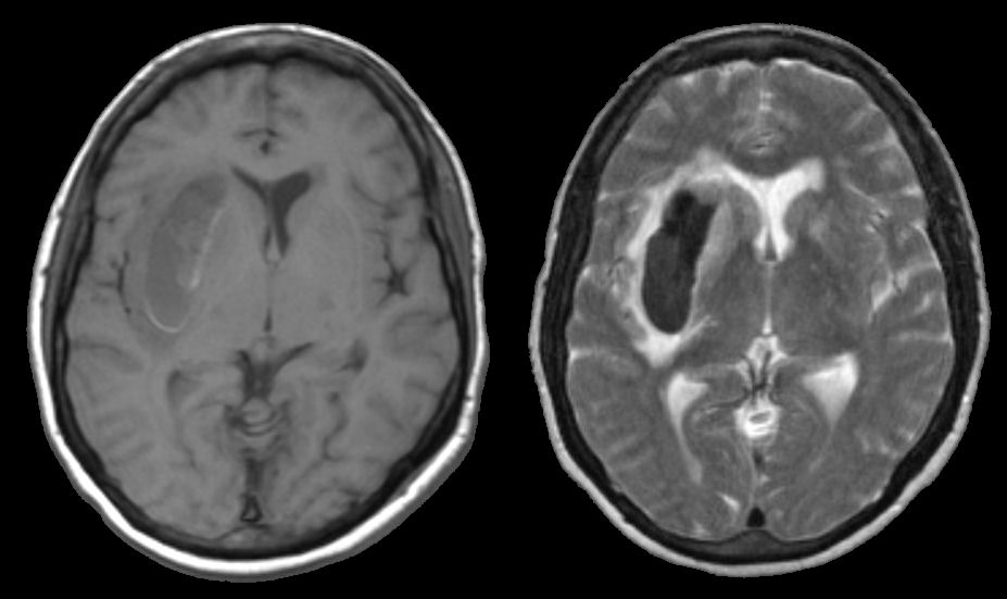
Axial MRI scans: (Left) T1-weighted; (Right) T2-weighted. Note on T1, there is an abnormality that is isointense in the right basal ganglia. The same area on T2 is dark with a surrounding bright signal. This is the characteristic picture of an acute (approximately 3 days old) hemorrhage on MRI. In the acute stage, intracellular deoxyhemoglobin is dark on both T1 and T2. As the deoxyhemoglobin changes to intracellualr methemoglobin, the signal because bright on T1 but remains dark on T2. Thus, when this transition occurs, there is a time where the signal is isointense on T1, as is seen here. Looking closely at the T1 scan, there is a faint rim of increase signal. This represents the early formation of intracellular methemoglobin. The findings of blood on MRI are complex and depend on timing. To learn more, review the powerpoint slide show, Blood on MRI: Time-dependent Changes. In this case, the hemorrhage was due to hypertension.
Revised05/02/06.
The Electronic Curriculum is copyrighted 1998, Case Western
Reserve University
School of Medicine.