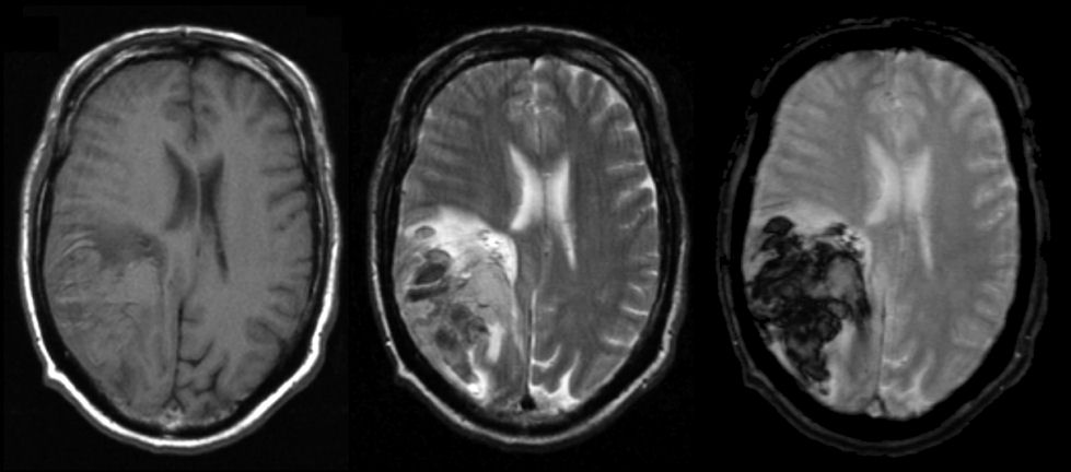
An 74 year old man presented with headache, a left visual field loss and numbness on the left side.
![]()
![]()
![]()
![]()
Axial MRI scans: (Left) T1-weighted; (Middle) T2-weighted; (Right) Gradient Echo. Note on T1, there is an abnormality that is isointense in the right parietal lobe. The same area on T2 is isotense or dark with a surrounding bright signal. The dark signal on T2 represents deoxyhemoglobin whereas the isotense signal is oxyhemoglobin. The surrounding bright signal is vasogenic edema. On gradient echo, the lesion is very dark - gradient echo is very sensitive in detecting hemoglobin. This is the characteristic picture of an hyperacute hemorrhage changing to an acute (approximately 3 days old) hemorrhage on MRI. The findings of blood on MRI are complex and depend on timing. To learn more, review the powerpoint slide show, Blood on MRI: Time-dependent Changes. In this case, the hemorrhage was due to hypertension.
Revised05/20/06.
The Electronic Curriculum is copyrighted 1998, Case Western
Reserve University
School of Medicine.