A 26 year old man was agitated and confused following a motor vehicle accident.
![]()
![]()
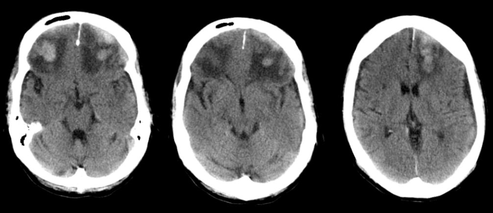
Axial CT scans of the brain. Note the area of traumatic contusion (i.e., hemorrhage and surrounding edema) in the both frontal lobes. There is also some mass effect on the anterior horns of the lateral ventricles. The frontal poles are common locations for cerebral contusions following head injury (i.e., the head stops but the brain keeps moving and strikes the inner skull).
Revised
05/20/06.
The Electronic Curriculum is copyrighted 1998, Case Western Reserve University
School of Medicine.