A 44 year old woman was in coma following a motor vehicle accident.
![]()
![]()
![]()
![]()
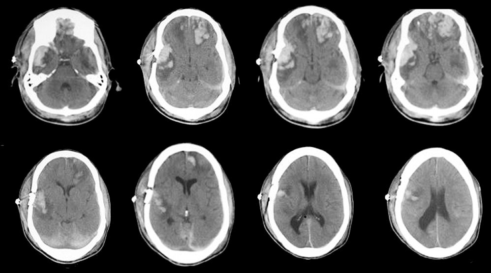
Axial CT scans of the brain. Note the large area of traumatic contusion (i.e., hemorrhage and surrounding edema) in the frontal lobes (left larger than right) and the right temporal lobe. Subarachnoid blood is also present, and most easily seen over the tentorium posteriorly. The frontal and temporal lobe tips are common locations for cerebral contusions following head injury (i.e., the head stops but the brain keeps moving and strikes the inner skull, making contact first in the frontal and temporal tips).
Revised
05/06/06.
The Electronic Curriculum is copyrighted 1998, Case Western Reserve University
School of Medicine.