A 42 year-old man was drowsy and confused following a hypotensive episode. Within a day, his neurologic condition markedly deteriorated and he fell into coma.
![]()
![]()
![]()
![]()
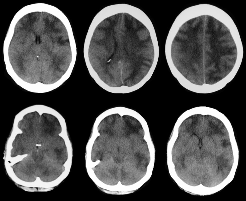
CT scan of the brain. Note the diffuse cerebral edema, most prominently seen in the white matter. No sulci are seen over the convexities and the cisterns around the brainstem have been effaced. This picture is most often seen following trauma, hypoxia or CNS infection, especially meningitis. Diffuse edema leads to increased intracranial pressure followed by decreased brain perfusion and then brain death.
Revised
04/25/06.
The Electronic Curriculum is copyrighted 1998, Case Western Reserve University
School of Medicine.