A 52 year-old woman presented with headaches, confusion and multiple strokes over the past 2 months.
![]()
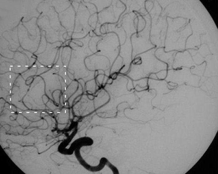
Cerebral Angiogram - Left Internal Carotid Artery injection - note the carotid artery is widely patent. However, upon close inspection of the distal branches, note the beaded appearance of the vessel. Normally, arteries taper as they travel more distally. This appearance is consistent with cerebral vasculitis, with can occur as an isolated syndrome or part of a more widespread system vasculitis. Often, brain and meningeal biopsy is needed to confirm the diagnosis. Cerebral vasculitis often presents with an encephalopathy and superimposed focal deficits from multiple ischemic strokes. The syndrome can be confused with embolic infarctions and arteriosclerotic intracranial disease, the latter which is more prominent in Asian and African-Americans rather than Caucasians.
Revised
04/25/06.
The Electronic Curriculum is copyrighted 1998, Case Western Reserve University
School of Medicine.