A 62 year-old man presented with progressive back and leg pain. His examination showed absent ankle reflexes and distal sensory loss.
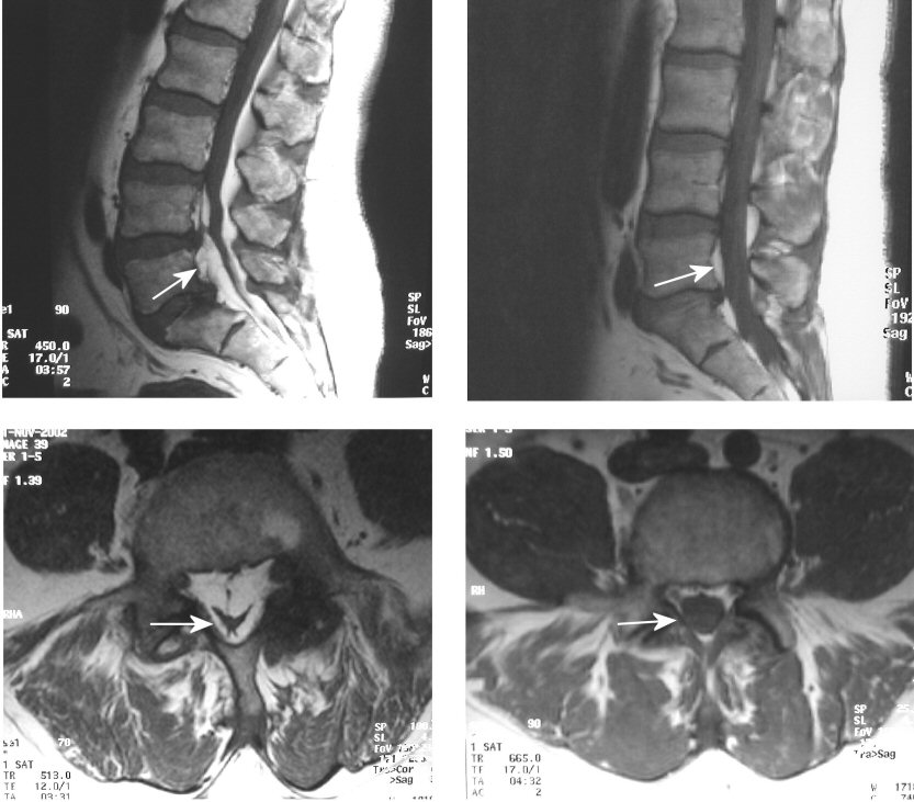
Axial T1-weighted MRI scan (Top - Sagittal; Bottom - Axial; Left - Patient, Right - Normal): Note that in T1-weighted images, fat appears bright. Normally, there is a small amount of epidural fat surrounding the thecal sack (right images). However, in some cases, the adipose becomes increased and compresses the thecal sac and exiting nerve roots (left images). The thecal sac may develop a stellate configuration (lower left). Epidural lipomatosis is an unusual condition where the amount of epidural fat becomes symptomatic. It can mimic disk disease and spinal stenosis.
Revised
06/10/04.
The Electronic Curriculum is copyrighted 1998, Case Western Reserve University
School of Medicine.