A 47 year-old man underwent routine lumbar laminectomy for a herniated disk. Following the surgery, he developed progressive weakness and then frank paraplegia and urinary incontinence. On examination, he had a sensory level at L1 and no reflexes in the legs.
![]()
![]()
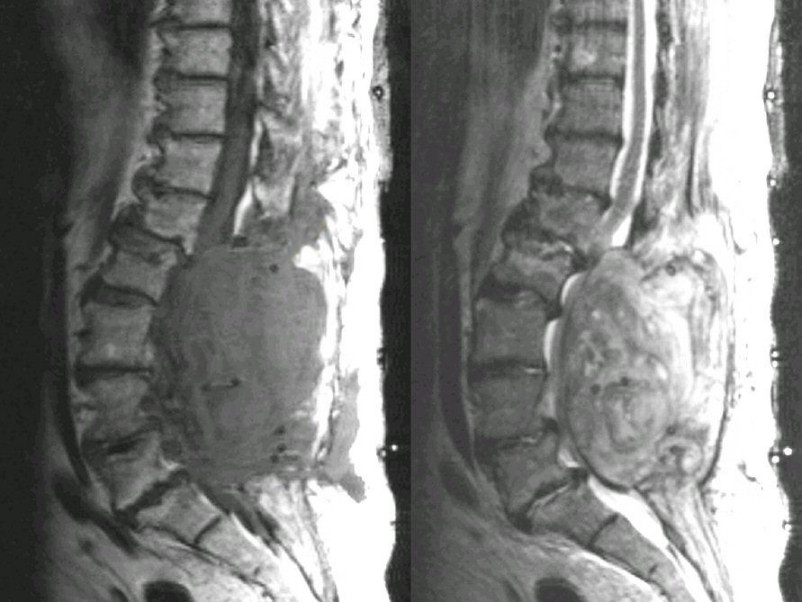
Sagittal MRI of the Thoracic Spine (Left - T1 weighted, Right - T2 weighted). Note the large lesion extradural lesion which is compressing the thecal sac from L2 through S1. It is iso-intense on T1 and slightly bright on T2. This is an acute epidural hematoma. Subsequently, the patient underwent emergency decompressive surgery
Revised
05/09/06.
The Electronic Curriculum is copyrighted 1998, Case Western Reserve University
School of Medicine.