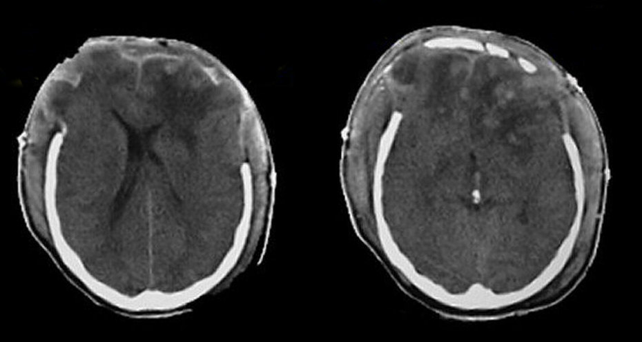
A 35 year old man suffered a head injury while participating in recreational activities. Bilateral craniectomies were done emergently for massive bilateral subdural and epidural hematomas. Post-operatively, he remained stuporous and a repeat CT was performed.
![]()
Axial CT scan: Note the areas of edema and contusion in both frontal lobs. Also note herniation of brain tissue through the craniectomy sites. In this case, the craniectomy defects allowed a path of herniation and potentially avoided transtentorial herniation.
Revised
05/07/06.
The Electronic Curriculum is copyrighted 1998, Case Western Reserve University
School of Medicine.