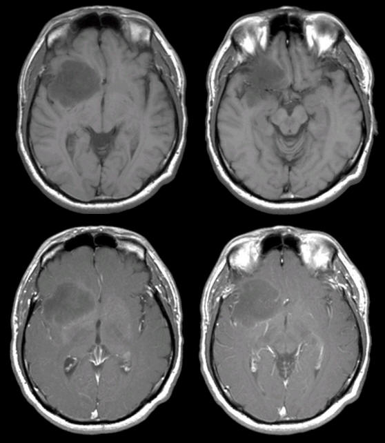
A 36 year-old man developed headaches and a change in personality over 6 weeks.
![]()
![]()
(Top Row) T1-weighted MRI axial scans; (Bottom Row) T1 weighted axial scans with Gadolinium. Note the large low density mass in the right inferior frontal lobe extending into the adjacent temporal lobe and basal ganglia. There is no contrast enhancement (note the contrast in the arteries and sinuses). Also, note that there is very little mass effect. This is a low grade glioma. Low-grade astrocytomas make up 15% of all primary intracranial brain tumors, and usually occur in young adults. Although biopsy is required to make a definitive diagnosis, the lack of contrast enhancement favors the diagnosis of a low grade glioma rather than a glioblastoma.
Revised
05/08/06.
The Electronic Curriculum is copyrighted 1998, Case Western Reserve University
School of Medicine.