|
Hydrocephalus
Alan R. Cohen, M.D.
Department of Neurological Surgery
Rainbow Babies and Children’s Hospital
Telephone: 216-844-5741
E-mail:
alan.cohen@uhhs.com
Definitions:
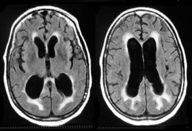
Hydrocephalus (Greek: hydro-water, kefale-head) has
been variably defined as an anatomic, radiographic,
physiologic or clinical phenomenon. For practical
purposes, hydrocephalus is best thought of as a
pathological accumulation of intracranial cerebrospinal
fluid (CSF), usually not always within the cerebral
ventricles.
Hydrocephalus can be classified as obstructive, that
is, associated with impairment in the circulation or
absorption of CSF, or non-obstructive, that is, a
relative enlargement of the ventricular system and CSF
spaces due to a loss of brain (ex vacuo hydrocephalus).
Hydrocephalus may also be congenital or acquired.
Obstructive hydrocephalus can be further divided into
communicating hydrocephalus, that is, obstructive
hydrocephalus that results from a blockage outside of
the ventricular system such that the ventricular fluid
is in communication with the subarachnoid space, and
non-communicating hydrocephalus, that is obstructive
hydrocephalus that results from a blockage within the
ventricular system and thus prevents communication with
the subarachnoid space.
Communicating hydrocephalus is more common than
non-communicating hydrocephalus. Examples of
communicating hydrocephalus include post-meningitic or
post-hemorrhagic hydrocephalus. Any process that scars
the subarachnoid space can lead to communicating
hydrocephalus. Examples of non-communicating
hydrocephalus include aqueductal stenosis or ventricular
tumors that obstruct the ventricles (e.g. pineal tumors
that block the aqueduct) and trap the CSF made proximal
to the obstruction.
Whereas hydrocephalus is usually a progressive
disorder marked by characteristic symptoms and signs,
sometimes the clinical features of hydrocephalus may
stabilize. The term arrested hydrocephalus is used to
describe a condition which is both non-progressive and
asymptomatic, while the term compensated hydrocephalus
is used to describe a condition which is
non-progressive, but symptomatic.
Epidemiology:
The prevalence of hydrocephalus in the general
population is unknown because the condition may occur in
isolation or in association with other congenital or
acquired disorders. As an isolated condition, the
incidence of hydrocephalus is approximately 1 to 1.5 per
1,000 births in the United States. When associated with
other disorders, the incidence of hydrocephalus is 3 to
4 per 1000 live births. One can estimate the prevalence
of hydrocephalus by looking at the treatment of this
condition. In the United States there are approximately
125,000 patients with CSF shunts. Each year there are
about 50,000 shunt operations.
History:
In 1949 Drs. Frank Nulsen and Eugene Spitz of the
University of Pennsylvania performed a landmark
operation to manage an infant with advanced
communicating hydrocephalus. They used a rubber tube
containing a one way stainless steel ball valve to
divert CSF from the enlarged cerebral ventricles to the
jugular vein. The surgery was successful and the
infant’s hydrocephalus came under control. As of this
writing, the patient is still alive and employed. The
operation of Nulsen and Spitz marked the first
successful implantation of a valved ventricular shunt,
and became a turning point in the surgical treatment of
hydrocephalus. Hydrocephalus was no longer a fatal
disorder, but was treatable such that infants and
children could survive to become adults with useful,
productive lives. There is no doubt that the
introduction of ventricular shunting in the second half
of the last century has had a revolutionary impact upon
the lives of patients with hydrocephalus. Of note, Frank
Nulsen went on to become the Chairmen of the Department
of Neurological Surgery at Case Western Reserve
University. He died several years ago.
In spite of ventricular shunting procedures, the
treatment of hydrocephalus remains a treacherous
undertaking for the neurosurgeon today. Shunt insertion,
usually one of the simplest of all neurosurgical
procedures, may also be one of the most complex. Shunts
are foreign bodies which can be troublesome to manage
because of a host of complications. Such complications
include malfunction (related to either underdrainage or
overdrainage to CSF) as well as infection. In fact,
ventricular shunting is associated with a higher
complication rate than any other commonly performed
neurosurgical procedure.
The evolution of the current management of
hydrocephalus is a remarkable story, but the powerful
impact of ventricular shunting should not leave
neurosurgeons with a sense of complacency. In a sense,
we have found a solution to a problem before we fully
understand its cause. The solution is a good one, but
far from ideal. The more we learn about the
pathophysiology of hydrocephalus, the greater the chance
of finding a better solution to the problem. As we look
back on many of the seemingly naïve ideas espounded in
the past, one cannot help but wonder how primitive
today’s “state of the art” treatment will be considered
by future neurosurgeons. The purpose of this lecture is
to review the evolution of the medical and surgical
management of hydrocephalus, and to discuss the benefits
and pitfalls of today’s state of the art in therapy.
Some important historical landmarks are listed below:
Hippocrates (5th century B.C.): Recognized that the
head could swell in response to an accumulation of water
within it. He felt that hydrocephalus was the result of
chronic epilepsy, and that water accumulated when the
diseased brain corroded and began to melt.
Claudius Galen (130-200 AD): Understood that the
brain was immersed in CSF. Expanded on the work of
Hippocrates. He provided a description of the choroid
plexus, but incorrectly believed that it secreted a
“psychic pneuma” which drained into the cribriform plate
and pituitary gland.
Thomas Willis (1621-1675): Remembered for his
description of the circular arterial anastomosis at the
base of the brain which bears his name. He was the first
to recognize that CSF was secreted by the choroid plexus
and drained into the venous system. Willis was less
accurate about the actual site of CSF absorption: he
believed this occurred within the nose after CSF had
passed through the cribriform plate.
Antonius Pacchioni (1701): Described the bodies that
today bear his name. He was able to provide a beautiful
illustration of the granulations but, as was typical of
many of the early investigations, not all his
observations were accurate. Pacchioni incorrectly
believed that the arachnoidal granulations were the
source of CSF production rather than the site of
absorption.
Key and Retzius (1875): Provided a lucid description
of the pathways of CSF movement from production to
reabsorption into the venous system. Once the CSF
circulation had been better worked out, a number of
investigators began to try innovative techniques to
control hydrocephalus.
Heinrich Quincke (1891): Described lumbar puncture as
a method of treatment for hydrocephalus, and recommended
enlarging the dural opening by moving the needle about.
Kausch (1908): First to place a ventriculoperitoneal
shunt. He used a rubber tube to connect the lateral
ventricle with the peritoneal space. Unfortunately, the
patient died on the day following surgery. Kausch felt
this was related to overdrainage of CSF.
Victor Darwin Lespinasse (1910): Attempted to treat
hydrocephalus by coagulating the choroid plexus, having
first cannulated the ventricles in two children using a
cystoscope. Although the event received little
attention, it marked not only the first choroid plexus
coagulation, but also the first use of an endoscope for
a neurosurgical procedure.
Walter Dandy (1918): Attempted to coagulate and
avulse the choroid plexus endoscopically, with limited
success. Several investigators have described
coagulation of the choroid plexus subsequently, but
these procedures have been largely abandoned by virtue
of their limited success in the treatment of
hydrocephalus.
W. Jason Mixter (1923): First endoscopic third
ventriculostomy. Mixter practiced the technique on a
cadaver and then used a small urethroscope and flexible
sound to fenestrate the floor of the third ventricle in
a 9 month old hydrocephalic infant, permitting egress of
CSF from the obstructed ventricular system into the
interpeduncular cistern. The procedure was successful,
and is a treatment for non-communicating hydrocephalus.
Nulsen and Spitz (1949): First successful valved
shunt insertion.
John Holter (1950’s): The initial ball valve used by
Nulsen and Spitz was primitive and ineffective. A better
“slit” valve was developed by Holter in the mid 1950s.
Holter was a blue collar worker from Pennsylvania whose
son, Casey, was born with a myelomeningocele and
hydrocephalus. Casey was shunted by Spitz using a
conventional system, but this shunt functioned poorly.
Working in a machine shop, often at night, Holter
developed the slit valve to treat his own son’s
hydrocephalus. This valve was used successfully by Spitz
to treat Casey, but unfortunately Casey later died of
complications of hydrocephalus. Holter’s slit valve
became one of the most widely used shunt vavles
throughout the world. He stumbled upon this discovery
quite fortuitously, watching nurses administer
intravenous medication to his infant son by placing a
needle through a rubber diaphragm on a piece of T-tubing
in the intravenous line, and noting that there was no
reflux of fluid.
The high complication rate for ventriculovascular
shunts and their requirement for frequent revision, led
ultimately to the development of the
ventriculoperitoneal shunt, which remains the surgical
standard for the treatment of hydrocephalus today.
Non-Surgical Treatment of Hydrocephalus:
Medical treatment of hydrocephalus is not very
effective, and is usually viewed as a temporizing
measure.
1. Medications that decrease CSF production and
reduce intracranial pressure:
acetazolamide
furosemide
2. Medications that reduce intracranial pressure:
mannitol
glycerol
urea
isosorbide
3. Medications that promote CSF absorption (not commonly
used):
hyaluronidase
heparin
urokinase
4. Intermittent CSF removal (e.g. serial lumbar
punctures)
Surgical treatment of hydrocephalus:
A variety of CSF shunt systems are currently
available. No single shunt has been clearly shown to be
superior to the rest, and thus the surgeon’s familiarity
with the shunt equipment is an important factor in its
selection. The consistent use of the same shunt system
helps to optimize the surgeon’s technical proficiency
and facilitates the process of shunt revisions.
The equipment for CSF diversionary shunting includes
a proximal shunt catheter, valve, distal shunt catheter,
and sometimes other components such as a device to
prevent siphoning, an on/off switch or telemetric
sensor. Shunt systems may exist as integral units (no
connectors) or as separate parts requiring assembly.
The proximal catheter is the portion of a shunt
system which is placed into the CSF space before the
site of obstruction. In ventricular shunt systems, the
proximal catheter is most commonly placed in the lateral
ventricle. When the subarachnoid space is shunted, the
proximal catheter is most commonly inserted into the
lumbar thecal sac. Proximal shunt catheters,
particularly ventricular catheters, should incorporate
several features in their design to ensure optimal
function. Some ventricular catheters include a
subcutaneous reservoir. The catheters may be straight or
right-angled. Right-angled catheters allow easier
fixation to the distal shunt system. The reservoir
should be palpable underneath the scalp and allow
percutaneous access to the CSF for sampling, pressure
measurement, and introduction of medication or contrast
agents.
Shunt valves come in several varieties. All produce
unidirectional flow of CSF. Most valves are pressure
regulated, that is, they respond to a differential
pressure gradient across the valve. A differential
pressure is generated either by an increase in pressure
upstream or a decrease in pressure downstream. The ideal
valve would drain only the excess CSF produced by each
individual patient which could not be reabsorbed. Such a
valve does not yet exist.
Ventricular shunt valves may be placed proximally or
distally along the shunt system. The majority of shunt
valves are proximal valves which are seated just distal
to the ventricular catheter. Distal valves of the slit
type are effective but have two major disadvantages: 1)
they are associated with a higher rate of distal shunt
malfunction and 2) they are more difficult to replace at
the time of shunt revision. Examples of proximal
differential pressure valves include the slit valve,
ball-in-cone valve, diaphragm valve, and miter valve.
Shunt Complications:
In spite of their dramatic ability to control the
symptoms and signs of hydrocephalus, ventricular shunts
are foreign bodies associated with a myriad of
complications. One must be familiar with the full
spectrum of complications that can follow shunt
placement because only some occur immediately and many
occur over the long-term. At times the rationale for
placement of a shunt presumes that because the surgery
is simple, “a shunt won’t hurt and might help”. While
the latter statement is often correct, the former is a
dangerous misconception. Complications that accompany
shunt placement are generally related to malfunction and
infection. Shunt malfunction may result from either
underdrainage or overdrainage of CSF.
Shunt malfunction from Underdrainage of CSF
Underdrainage of CSF occurs if the shunt system
becomes obstructed or disconnected. Most commonly, this
occurs as the result of an obstruction of the
ventricular catheter. This obstruction may be the result
of initial misplacement of the ventricular catheter or a
migration of the catheter into the subependymal tissue
or choroid plexus as a result of collapse of the
ventricles, growth or the head, or movement of the
entire shunt system. Even a catheter properly placed
within the ventricles may become occluded by choroid
plexus or tissue debris. Occlusion of a catheter as a
result of debris from an immune response has also been
described. The shunt valve may stick or become occluded
and malfunction. The distal shunt is also a site which
can become occluded, although this is more apt to occur
if a distal slit valve apparatus is used, and less
likely to occur if open ended peritoneal tubing is used.
Underdrainage of CSF may also result from a
disconnection with the shunt system. Disconnections tend
to occur at connector sites, particularly when
connectors have been placed along the shunt tract to
bridge gaps at the time of previous shunt
disconnections. Disconnections may occur anywhere along
the shunt system. When shunts have been in place for
long periods of time the tubing may become brittle and
even calcify, making it more prone to fracture.
Disconnections are common during growth spurts and tend
to occur at areas of movement such as the neck or at
areas subject to pressure such as the region overlying
the clavicle. Disconnections have become less common
with newer systems that contain fewer connector sites.
Underdrainage may also be the result of loculation
within the ventricular system. Loculated ventricles may
occur following hemorrhage or infection. Occlusion of
the foramen of Monro can create a trapped lateral
ventricle. Multiple septations may occur within the
ventricles, and a single shunt may be ineffective in
draining these isolated fluid collections.
Ventriculoscopic techniques can be used to fenestrate
the septum pellucidum as well as the walls of loculated
cysts. This allows simplification of shunt systems and
in some cases the need for a shunt may be eliminated
(e.g. after septostomy to bypass a blocked foramen of
Monro, a trapped lateral ventricle may drain through
normal pathways on the other side).
Ventriculoatrial shunts have a high rate of
malfunction. These shunts require frequent revision
because the distal end migrates out of the cardiac
atrium during growth of the child. Ventriculoatrial
shunts are also subject to problems of vascular or
cardiac perforation, embolism, and an immune-mediated
glomerulonephritis.
Shunt Malfunction from Overdrainage of CSF
All extracranial CSF shunts (ventriculo-peritoneal,
-pleural and -atrial) are subject to malfunction from
overdrainage. In each of these shunts there may be
markedly negative pressures generated by the hydrostatic
column of fluid in the tubing distal to the shunt valve
when the patient assumes the upright position. The
problem of overdrainage of CSF is common to the
differential pressure valves, as the negative
hydrostatic pressure generated in the upright position
can overcome the effect of even the “high pressure”
valves.
The most common symptom from overdrainage is
headache. This low pressure headache must be
distinguished from the high pressure headache due to CSF
underdrainage. Overdrainage headache is worse in the
upright position and improved when the patient is
recumbent. It is often transient and will abate if the
patient is allowed to adjust to the upright position
slowly. Occasionally, upgrading the valve or use of a
device to prevent siphoning is necessary.
When headaches are persistent one must consider the
possibility of a more dangerous condition such as a
subdural hematoma or hygroma. This can be a very
difficulty problem to treat, particularly in patients
who are shunt dependent. Burr hole drainage of the
subdural collection, or subdural to peritoneal shunting
with a valveless system can be utilized.
The slit-ventricle syndrome is a condition
characterized by intermittent headache and symptoms
suggestive of shunt malfunction from underdrainage of
CSF that occurs in children with small slit-like
ventricles. When these children are symptomatic, the
intracranial pressure is elevated. These children have
usually been shunted as infants and often have small
heads. The shunt valve refills slowly. There may be
papilledema or cranial nerve abnormalities and,
occasionally, hypertension and bradycardia. Many authors
regard the symptoms and signs of the slit-ventricle
syndrome to be manifestations of intermittent shunt
obstruction due to small ventricular size occurring in a
brain that lacks compliance, perhaps due to a diminished
craniocephalic ratio. Although many children with
indwelling ventricular shunts have small or slit-like
ventricles, the slit-ventricle syndrome is relatively
rare, occurring in 1-5% of infants shunted for
hydrocephalus.
Shunt Infection
Shunt infections make up a substantial percentage of
the complications that accompany shunt placement.
Despite a markedly improved trend in infection rate over
the past two decades, shunt infections are still
reported in 2-10% of cases. The presence of
ventriculitis has been shown to adversely affect
intelligence. In one study, the average IQ of shunted
patients having had ventriculitis was 72 as compared to
95 in shunted patients not having had ventriculitis.
Staphylococcus epidermidis is the most common
organism to infect ventricular shunts. The majority of
shunt infections occur within two months of shunt
insertion. For this reason it is presumed that the
infection is the result of intraoperative contamination.
It is interesting that cultures of the skin of shunt
recipients do not consistently match subsequent cultures
of infected shunts. This suggests either that the source
of seeding can be from an area other than the
recipient’s skin, such as the nasopharynx, or from the
skin of operating room personnel.
Alternative Operative Approaches to Hydrocephalus:
Endoscopic Third Ventriculostomy
Percutaneous endoscopic third ventriculostomy (ETV)
has been repopularized as a technique for bypassing an
obstruction at the aqueduct of Sylvius or fourth
ventricle in non-communicating hydrocephalus. It has
gained appeal as the result of advances in stereotactic
and endoscopic technology, and in some cases may
eliminate the need for ventricular shunting and its
associated risks.
ETV is performed through a standard coronal burr hole
approach. As mall endoscope is guided into the lateral
ventricle, then through the foramen of Monro into the
third ventricle. A probe is used to puncture the floor
of the third ventricle anterior to the mammillary
bodies. The fenestration is enlarged using a small
balloon catheter. All instruments are introduced through
working channels in the thin endoscope sheath, so the
procedure is “minimally invasive”. It works by allowing
CSF to exit the ventricular system and then circulate
normally in the subarachnoid space. ETV is thus an
effective treatment for non-communicating hydrocephalus
e.g. aqueductal stenosis. It is not effective for
communicating hydrocephalus, because the obstruction in
this condition is further downstream.
ETV works best in patients with acquired or
late-onset presentation of aqueductal stenosis.
Presumably, this is because these patients have already
developed adequate absorptive pathways distal to the
acquired aqueductal obstruction. In such patients, third
ventriculsotomy is simply a means of bypassing an
obstruction at the aqueduct of Sylvius. Careful patient
selection is essential for successful third
ventriculostomy.
Choroid Plexus Coagulation
Coagulation of choroid plexus has been utilized as a
treatment for hydrocephalus with generally
unsatisfactory results. The aim of treatment is to
decrease CSF pressure by reduction CSF production. Since
not all CSF is produced by the choroid plexus, there is
a theoretical limit to the efficacy of this procedure.
The idea of removing or coagulating choroid plexus as
a treatment for hydrocephalus dates back to the turn of
the century. Early procedures met with limited success,
but subsequent investigators reported improved results
and lower morbidity and mortality rates. The efficacy of
choroid plexus coagulation is difficult to assess
because there are no controlled comparisons with
ventricular shunting procedures, and authors use
variable criteria to judge success of the procedure. In
general, although it is now technically feasible,
choroid plexus coagulation has been largely abandoned as
ineffective as a means of controlling hydrocephalus.
Summary:
Introduction of the valved ventricular shunt in the
middle of the 20th century has revolutionized the
management of hydrocephalus. Infants, children and
adults with a once fatal disorder can now enjoy useful
and productive lives. Nevertheless, ventricular shunts
remain troublesome and are subject to numerous
complications. A re-examination of the pathophysiology
of hydrocephalus may lead to improved methods for its
treatment the optimal treatment would combine improved
efficacy with reduced complications. |