An 55 year old man presented with headache, vomiting and a left hemiparesis.
![]()
![]()
![]()
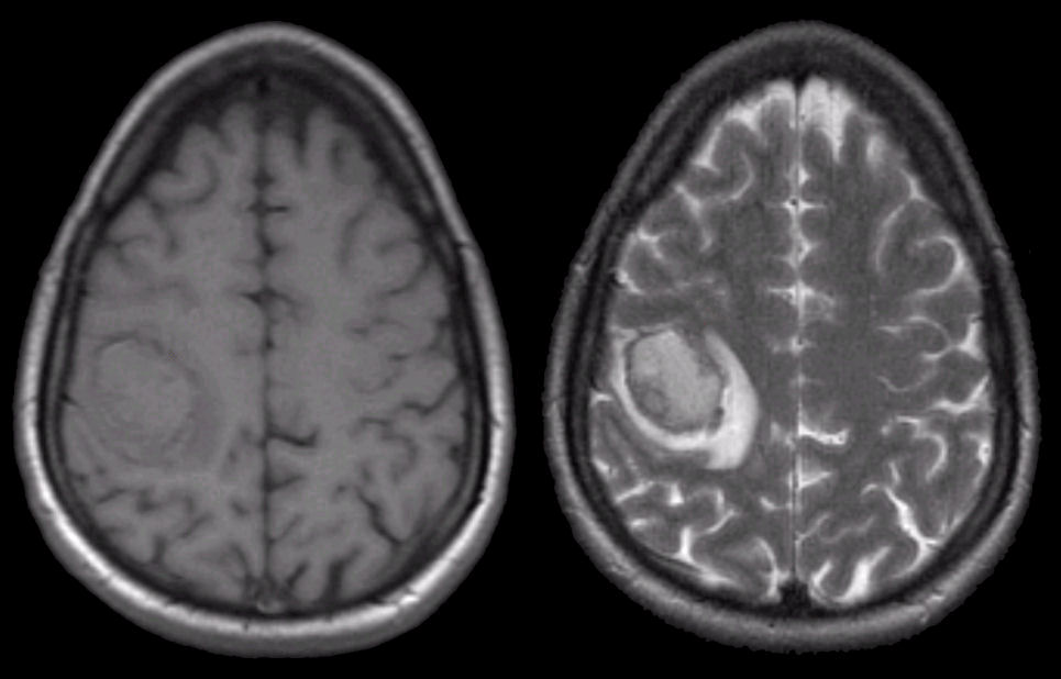
Axial MRI scans: (Left) T1-weighted; (Right) T2-weighted. Note on T1, there is an abnormality that is relatively isointense in the right posterior frontal lobe. The same area on T2 is isointense / hyperintense with a surrounding bright signal. This is the characteristic picture of a hyperacute (approximately 1 day old) hemorrhage on MRI. In the hyperacute stage, intracellular oxyhemoglobin is isointense / hypointense on T1 and isointense / hyperintense on T2 . Surrounding the lesion on T2 is a bright signal consistent with vasogenic edema. The findings of blood on MRI are complex and depend on timing. To learn more, review the powerpoint slide show, Blood on MRI: Time-dependent Changes. In this case, the hemorrhage was due to hypertension.
Revised
05/02/06.
The Electronic Curriculum is copyrighted 1998, Case Western
Reserve University
School of Medicine.