A 64 year-old man with poorly controlled hypertension had a remote history of weakness involving the left face and body.
![]()
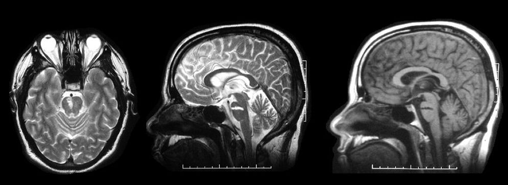
MRI axial images - (Left) T2 (axial at the level of the upper pons); (Middle) T2 (mid-sagittal); (Right) T1 (mid-sagittal): Note the well-demarcated ischemic stroke in the region of the basis pontis on the right. This type of stroke is caused by occlusion of the deep perforating blood vessels (also known as small vessel disease or lacunar strokes). Small vessel disease is most commonly associated with hypertension and diabetes. There are several classic lacunar syndromes, including pure motor hemiparesis, ataxic hemiparesis, clumsy hand-dysarthria (lesions either in the internal capsule or basis pontis) and pure sensory stroke (lesion in the thalamus). The well demarcated lesion of the T1 demonstrates that the lesion is chronic.
Revised
04/23/06.
The Electronic Curriculum is copyrighted 1998, Case Western Reserve University
School of Medicine.