A 46 year old woman presented with left hemiplegia and headache. The previous medical history was significant for hypertension.
![]()
![]()
![]()
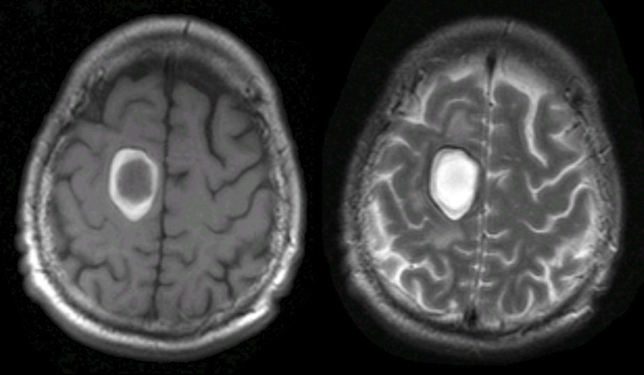
Axial MRI scans of the brain. (Left) T1 weighted; (Right) T2 weighted. Note the large hemorrhage in the right frontal lobe. On MRI, blood has a complex pattern, depending on whether the image is T1 or T2 weighted, and how long the hemorrhage has been present. The "white" on T1 and T2 represent extracelluar methemoglobin. Looking closely, there is a thin black surrounding rim. This is hemosiderin. These findings mark the hemorrhage as subacute.
Revised
05/04/06.
The Electronic Curriculum is copyrighted 1998, Case Western Reserve University
School of Medicine.