A 72 year-old man noted chronic low back pain, worst with standing and walking.
![]()
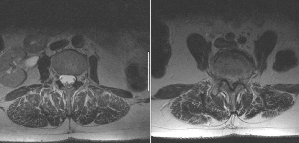
(Left) Axial T2-weighted MRI at L3; (Right) Axial T2-weighted MRI at the L4-5 disk level. Note the severe stenosis at the L4-L5 level with the complete absence of CSF. Contrast this to a normal level above at the L3 level and the normal amount of CSF surrounding the descending nerve roots of the cauda equina.
Revised
04/30/06.
The Electronic Curriculum is copyrighted 1998, Case Western Reserve University
School of Medicine.