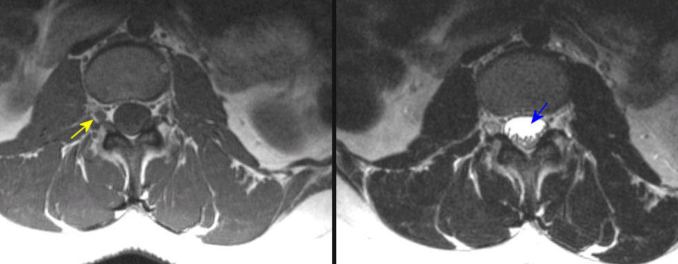
(Left)
T1-weighted image; (Right) T2-weighted image. Note on
the T1 scan, that the arrow points to an exiting nerve root in the intervertebral foramen. On the T2 image of the right, note that the CSF is
bright. The arrow points to the cross section of thecal sac where the descending nerve roots, known
as the cauda equina, are easily seen. Also note that on both T1 and T2, fat is
bright (look in the muscles) whereas fluid is dark on T1 and bright on T2.
Revised
09/13/05.
The Electronic Curriculum is copyrighted 1998, Case Western Reserve University
School of Medicine.