<% strPathPics = Session("strPathPicsL") imgBg = strPathPics + Session("strMedia") %> Normal Sagittal Cervical MRI

Normal Sagittal MRI scans of the Cervical Spine

C2 Vertebral Body C3 Vertebral Body C4 Vertebral Body C5 Vertebral Body C6 Vertebral Body C7 Vertebral Body T1 Vertebral Body T2 Vertebral Body T3 Vertebral Body T4 Vertebral Body Dens of C2 Ring of C1 Ring of C1 Spinal Cord Medulla Tonsils (cerebellum) Foruth Ventricle Formen of Magendie Cisterna Magna Pons Cerebellum Premedullary Cistern Prepontine Cistern Clivus Nasal Phargynx Oral Pharygnx Palate CSF (subarachnoid space) CSF (subaranchoid space) Posterior Bony Elements of the Spine Subcutaneous and adipose tissue Muscle Adipose Tissue C2-3 Disk C3-4 Disk C4-5 Disk C5-6 Disk C6-7 Disk C7-T1 Disk T1-2 Disk T2-3 Disk T3-4 Disk C2 Vertebral Body C3 Vertebral Body C4 Vertebral Body C5 Vertebral Body C6 Vertebral Body C7 Vertebral Body T1 Vertebral Body T2 Vertebral Body T3 Vertebral Body T4 Vertebral Body Dens of C2 Ring of C1 Ring of C1 Spinal Cord Medulla Tonsils (cerebellum) Pons Cerebellum Clivus Fourth Ventricle Foramen of Magendie Cisterna Magna Premedullary Cistern Prepontine Cistern CSF (subarachnoid space) CSF (subaranchoid space) Posterior Body Elements of the Spine Subcutaneous and adipose tissue Adipose Tissue Muscle Nasal Phargynx Oral Phargynx Palate C2-3 Disk C3-4 Disk C4-5 Disk C5-6 Disk C6-7 Disk C7-T1 Disk T1-2 Disk T2-3 Disk T3-4 Disk

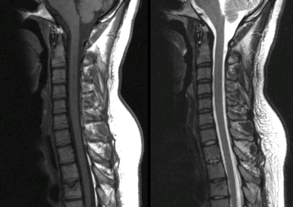
Left (T1-weighted image); Right (T2-weighted image). Note on the T2 image that CSF is bright and on the T1 image that CSF is dark.

Place the cursor over a structure to display the anatomy


Revised 05/09/06.
The Electronic Curriculum is copyrighted 1998,  Case Western Reserve University School of Medicine.