A 52 year-old woman presented with headaches, vertigo and falling.
![]()
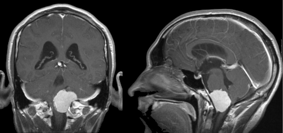
MRI Scans: (Left) T1-weighted coronal with gadolinium; (Right) T1-weighted sagittal with gadolinium Note the well demarcated mass which enhances with gadolinium, and nearly obliterates the medulla. On the sagittal scan, it is more easily seen that the mass is dural based and arising from the clivus. Also, note the cerebral aqueduct which is enlarged, reflecting the underlying obstructive hydrocephalus.
Revised
05/15/06.
The Electronic Curriculum is copyrighted 1998, Case Western Reserve University
School of Medicine.