A 65 year-old woman presented with headaches and mild sensory loss affecting the left side of her body.
![]()
![]()
![]()
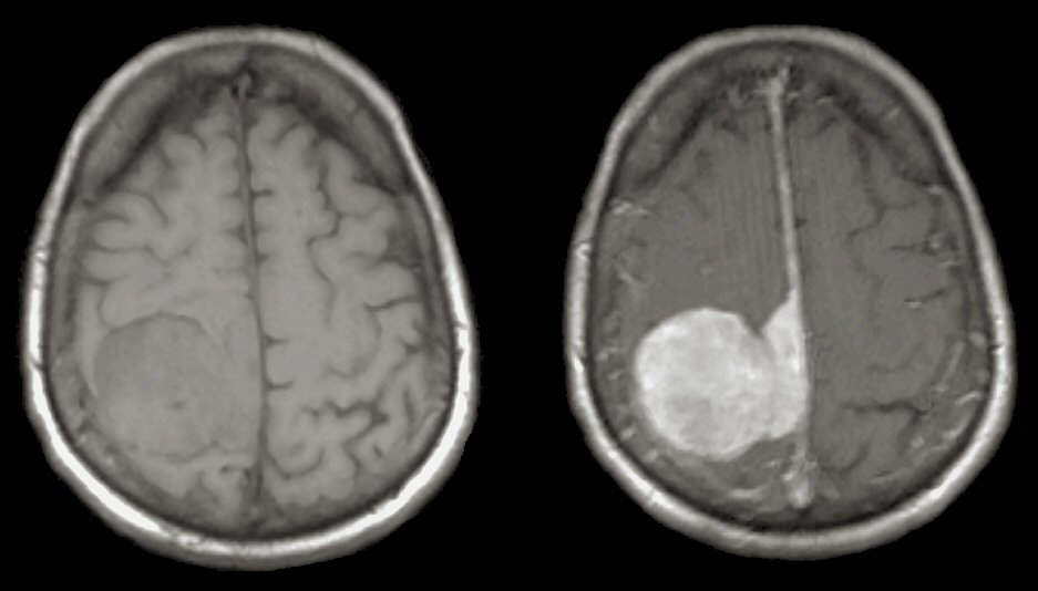
MRI Scans (Left: T1 weighted axial image; Right: T1-weighted image with gadolinium). Note the large, well demarcated mass which is attached to the falx in the midline with is expanding and compressing the adjacent parietal lobe. This is the typical appearance of a meningioma. They are typically benign histologically, and treatable with surgical resection if they are in an accessible location. In this location, if they grow large enough, they may grow into the superior sagittal sinus, that markedly increases the complexity and morbidity during surgical resection.
Revised
05/15/06.
The Electronic Curriculum is copyrighted 1998, Case Western Reserve University
School of Medicine.