(NL - normal level; CSF - cerebrospinal fluid; SAH - subaranchoid hemorrhage;
PMNs - polymorphonucleocytes)
Note the following important points:
1) CSF opening pressure needs to be measured in the recumbent position
with the patient's abdominal muscles relaxed; otherwise the pressure may be
factiously elevated (i.e., increased intra-abdominal pressure increases
intracranial pressure).
2) In early viral meningitis, there may be a brief phase of PMNs before a
lymphocytic predominance occurs.
3) CSF Glucose needs to be compared to serum glucose, preferably at the same
time (serum glucose 2-4 hours before the LP is typically acceptable). CSF
glucose typically lags behind serum glucose.
4) Low CSF Glucose (hypoglycorrhachia) is
classically seen in bacterial meningitis. However, a normal CSF glucose does not
rule out bacterial infection. In addition, hypoglycorrhachia can be seen in
chemical meningitis, inflammatory conditions (e.g., sarcoid), and subarachnoid
hemorrhage.
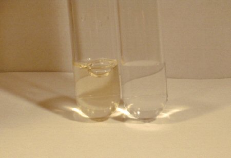
5) 20% of all LP are traumatic (presumably an epidural vein is entered). It
is essential to be able to differentiate a traumatic tap from a true SAH.
The following points help:
•
Compare the number of RBCs between the first and last
tubes. In a traumatic tap, the number typically decreases where it is
unchanged in SAH.
•
Measure the opening pressure. It is almost
always elevated in SAH and normal in a traumatic tap. |