|
Definition |
Immediate but transient loss of consciousness,
associated with short period of amnesia. |
Surface bruise of brain with varying degrees
of petechial hemorrhage, edema, and tissue destruction. |
|
Mechanism of Injury |
Transient electrophysiological dysfunction of
reticular activating system due to rotation of cerebral hemispheres
around a fixed brainstem. |
Deceleration of brain against the skull. Coup
lesion occurs at point of brain impact, contre-coup lesion occurs
when brain swings back. |
|
Clinical Signs |
Most patients are neurologically normal,
although brief convulsion, facial pallor, bradycardia, faintness
and sluggish pupillary reaction may occur.
Memory loss is usually limited to brief period
of time surrounding the injury; occasionally, weeks of memory loss
may occur.
Permanent neurological deficit following
concussion does not occur. |
Focal neurologic signs (e.g., hemiparesis, gaze
preference, etc.) and/or altered mental state. In severe lesions,
coma and extensor posturing.
Edema often follows as a secondary effect and
may put the patient at risk of herniation. |
|
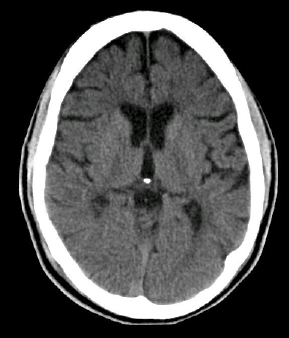
Imaging |
CT and MRI usually normal. |
CT: areas of contusion are often inhomogeneous
hyperdensities (i.e., blood) with surrounding edema and mass effect.
MRI: petechtial blood or frank contusion (MRI
appearance depends on sequence and timing). MRI may also be able to
visualize diffuse axonal shearing injury a few days later.
|