Neuroscience Clerkship


Note the subarachnoid blood (white) in the basal cisterns as well as in the left occipital horn of the lateral ventricle


Spontaneous Subarachnoid Hemorrhage:


Clinical Presentation,

Etiology, Complications

and Treatment


Definition


Subarachnoid hemorrhage (SAH) is the extravasation of blood into the subarachnoid space between the pial and arachnoid membranes.

Etiology

The most common causes of spontaneous SAH are rupture of saccular, or berry, aneurysm (80%); and rupture of arteriovenous malformation (AVM) (10%).

Other causes of non-traumatic SAH include: mycotic aneurysms, amyloid angiopathy, blood dyscrasias, fibromuscular dysplasia, idiopathic, infection, Moyamoya disease, neoplasm, and vasculitis (10%).

Epidemiology (Frequency, Age, Sex, Race)

Unlike other subcategories of stroke, the incidence of SAH has not decreased over time. However, since 1970, survival rates have improved. Annual incidence of aneurysmal SAH worldwide varies from 2-49 cases per 100,000 population, with the highest rates occurring in Japan and Finland. Annual incidence of aneurysmal SAH in the United States is 6-16 cases per 100,000 population, with approximately 30,000 episodes occurring each year. Incidence peaks at age 50.

There is a 3:2 female to male ratio; and a 2:1 black to white ratio. Incidence of SAH from aneurysmal and AVM rupture is significantly higher during pregnancy, especially the third trimester. SAH from aneurysmal rupture accounts for 6-25% of maternal deaths during pregnancy.
 

Aneurysm Pathophysiology

Aneurysms are specific to the intracranial arteries because their walls lack an external elastic lamina, they contain a very thin adventitia, and they lie unsupported in the subarachnoid space. The early precursors of aneurysms are small outpouchings through defects in the media of the arteries. These defects expand as a result of hydrostatic pressure from pulsatile blood flow and blood turbulence. A mature aneurysm has a paucity of media, replaced by connective tissue, and has diminished or absent elastic lamina. The probability of rupture is related to the tension on the aneurysm wall, as determined by the Law of La Place; thus, the rate of rupture is directly related to the size of the aneurysm.

Aneurysms with a diameter of 5 mm or less have a 2% risk of rupture

Aneurysms with a diameter of 6-10 mm have a 40% risk of rupture

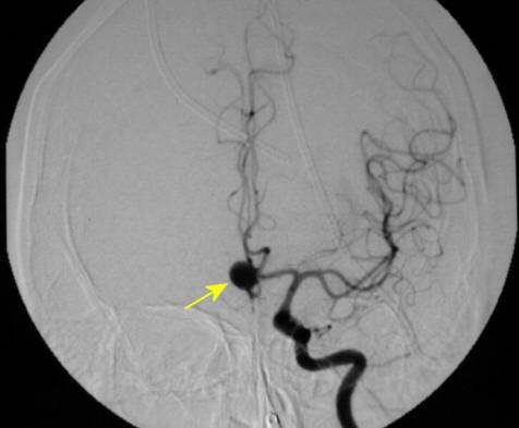
Aneurysms usually occur at arterial bifurcations

Aneurysms mostly arise from the anterior circulation of the Circle of Willis (85%)
 

The Most Common Sites of Aneurysms are:

Internal carotid artery/posterior communicating artery (41%)

Anterior communicating artery/anterior cerebral artery (34%)

Middle cerebral artery (20%)

Vertebral artery/basilar artery (4%)

Other arteries (1%)
 

Aneurysm Formation

Acquired factors associated with aneurysm formation include:

Atherosclerosis

Hypertension

Hemodynamic stress

Evidence supporting the association between congenital factors and aneurysm formation:

Clusters of familial occurrence (e.g., in Finland, the incidence of familial cerebral aneurysm is 10%)

Incidence of multiple aneurysms in patients with SAH (15-20%)

Association with specific congenital diseases (e.g., coarctation of the aorta, Marfan syndrome, Ehlers-Danlos syndrome, fibromuscular dysplasia, polycystic kidney disease)
 

Known Risk Factors for Aneurysm Rupture

Tobacco use

Alcohol abuse

Hypertension caused by cocaine and other stimulants

Large aneurysm size
 

Prodromal Signs and Symptoms

Prodromal signs and symptoms are the result of sentinel leaks, mass effect of aneurysm expansion, or emboli.

Sentinel, or "warning," leaks that produce minor blood leakage are reported to occur in 30-50% of aneurysmal SAHs; they do not usually occur in the setting of AVM.

Sentinel leaks produce sudden focal or generalized head pain that may be severe; they also produce nausea, vomiting, photophobia, malaise, or, less commonly, neck pain.

Sentinel headaches precede aneurysm rupture by a few hours to a few months, with a reported mean of 2 weeks prior to discovery of the SAH.

Mass effect of an expanding aneurysm has characteristic features based upon aneurysm location. The most classic is an ipsilateral 3rd nerve palsy from a Posterior Communicating Artery aneurysm.

Transient ischemic attacks can occur from emboli originating from intra-aneurysmal thrombus formation.


Signs and Symptoms of Aneurysmal Rupture

A sudden onset of severe headache ("thunderclap headache"), often described as the “worst headache of my life;” absence of headache in the setting of a ruptured aneurysm is rare.

Nuchal pain and rigidity, back pain, and bilateral leg pain secondary to meningeal irritation occurs in as many as 80% of patients, but may take several hours to manifest

A sudden loss of consciousness (LOC) occurs in half of patients at bleeding onset; it is usually transient, although 10% of patients are comatose for several days

Nausea and/or vomiting

Photophobia and/or visual disturbances

Seizures occur in 10-25% of patients, usually in the first few minutes after bleeding onset

Less severe hemorrhages may present with headache of moderate intensity, neck pain, and nonspecific symptoms

Approximately 30-40% of patients are at rest at the time of SAH
 

Physical exam
 

Blood pressure elevation is observed in about 50% of patients; blood pressure often becomes labile as ICP increases

Tachycardia often is present for several days after SAH

Temperature elevation, secondary to chemical meningitis from subarachnoid blood, is common after the fourth day following bleeding

Global depression of neurological function may be noted, including altered level of consciousness and confusional state in 25% of patients

Papilledema Subhyaloid hemorrhage Large Subhyaloid Hemorrhage Subhyaloid hemorrhage

Ophthalmologic examination may reveal subhyaloid retinal hemorrhages (small round hemorrhage, often with visible meniscus, near the head of the optic nerve) in 20-30% of patients and papilledema

Focal neurologic abnormalities, including hemiparesis, aphasia, hemineglect, cranial nerve palsies, and memory loss, are present in 25% of patients

There are no localizing signs in 40% of patients
 

Clinical grading scales:
    Hunt and Hess scale

Grade 1 - Asymptomatic or mild headache

Grade 2 - Moderate-to-severe headache, nuchal rigidity, and no neurological deficit other than possible cranial nerve palsy

Grade 3 - Mild alteration in mental status (confusion, lethargy), mild focal neurological deficit

Grade 4 - Stupor and/or hemiparesis

Grade 5 - Comatose and/or decerebrate rigidity
 

World Federation of Neurological Surgeons (WFNS) scale

Grade 1 - Glasgow Coma Score (GCS) of 15, motor deficit absent

Grade 2 - GCS of 13-14, motor deficit absent

Grade 3 - GCS of 13-14, motor deficit present

Grade 4 - GCS of 7-12, motor deficit absent or present

Grade 5 - GCS of 3-6, motor deficit absent or present

The Hunt and Hess and the WFNS grading scales have been shown to correlate well with patient outcome.


Workup (Lab, Imaging, and Procedures)


Serum chemistry panel, CBC, coagulation panel, blood type and screen, cardiac enzymes, EKG

Computed tomography (CT) head without contrast

CT has a sensitivity of 98% at 12 hours, 93% at 24 hours, 80% at 72 hours, and 50% at 1 week

Blood localized to the basal cisterns, the Sylvian fissure, or the intrahemispheric fissure may indicate the likely site of the aneurysm

Blood found lying over the convexities or within the superficial parenchyma of the brain often is indicative of AVM or mycotic aneurysm rupture

CT findings may be falsely negative in patients with small hemorrhages and in patients with severe anemia

If CT does not show SAH but clinical suspicion remains high, an LP must be performed

Lumbar puncture (LP)

Cerebrospinal fluid (CSF) is evaluated for the presence of red blood cells (RBCs) and xanthochromia

RBC number in the CSF after SAH can vary; elevated RBC number may also indicate a technically traumatic puncture

RBCs in the CSF remain consistently elevated in 2 sequential tubes or punctures in SAH, whereas the number of RBCs in a traumatic tap decreases over time

D-dimer assay has also been used to discriminate SAH from traumatic LP

Xanthochromia refers to a pink or yellow coloration of the CSF supernatant caused by the breakdown of RBCs and the subsequent release of heme pigments. Xanthochromia typically will not appear until 2-4 hours after bleeding onset. Xanthochromia is present 12 hours after bleeding onset and remains for approximately 2 weeks in nearly 90% of patients with SAH; it is present in 70% of patients at 3 weeks and 40% of patients at 4 weeks. Spectrophotometry is much more sensitive than the naked eye in detecting xanthochromia.

Xanthochromia (left) compared to normal CSF (right)

Above:  Xanthochromia refers to a pink or yellow coloration of the CSF supernatant (left tube) caused by the breakdown of RBCs and the subsequent release of heme pigments compared to normal (right tube).
Definitive Imaging


After the diagnosis of SAH, further imaging should be performed to characterize the source of the hemorrhage.

Cerebral angiography is the gold standard to assess for aneurysms. Both vertebrals and both carotids always need to be studied, as multiple aneurysms can occur in a patient. Angiography can define the aneurysm location, size, shape, as well as the orientation of the aneurysm dome and neck, presence of additional aneurysms (about 15-20% of patients have multiple aneurysms), and can help with operative planning.

Note the large Acomm aneurysm

Figure Above: Left Internal Carotid angiogram, demonstating a large Anterior Communicating Artery aneurysm.

Approximately 10-20% of patients with SAH diagnosed by CT and/or LP have negative angiographic findings. These patients may have an improved prognosis. In this group, a repeat angiogram is usually indicated in 3-4 weeks (note - rarely clot inside an aneurysm or vasospasm will mask an aneurysm on angiography.


Magnetic resonance angiography (MRA) is less sensitive than cerebral angiography at detecting small aneurysms; however, it is generally thought that MRA will eventually replace conventional cerebral angiography as it is non-invasive.

Computed tomography angiography (CTA) is beneficial in unstable patients who cannot undergo cerebral angiography or in emergent settings prior to operative interventions.

Magnetic resonance imaging (MRI) is also a useful tool; it can be used to diagnose and monitor unruptured cerebral aneurysms 5 mm or larger, to evaluate the degree of intramural thrombus in giant aneurysms, and to diagnose AVMs that are not detected by cerebral angiography.


Complications


Rebleeding

Approximately 6% of rebleeds occur within 24-48 hours of bleeding onset, then at a rate of 1.5% per day for the next 12-13 days, culminating in a two-week incidence of 20-30%.

Rebleeds in the first 2 days ("blow out" hemorrhages) are thought to be related to the unstable nature of the aneurysmal thrombus rather than lysis of the clot overlying the rupture site. The overall mortality rate from rebleeding is reported to be as high as 75%.

Vasospasm

Vasospasm is the delayed narrowing of large capacitance vessels at the base of the brain

Vasospasm is reported to occur in as many as 70% of patients with SAH and is clinically symptomatic in as many as 30% of patients; it most commonly occurs 4-14 days after bleeding onset

Vasospasm can lead to impaired cerebral autoregulation and may progress to cerebral ischemia and infarction (i.e., stroke)

Risk factors for vasospasm include larger volumes of blood in the subarachnoid space, clinically severe SAH, female sex, young age, and tobacco use

Conventional angiography is the definitive imaging study for vasospasm, although the diagnosis can be made reliably at the bedside using transcranial Doppler

If vasospasm becomes symptomatic, the use of hypertensive, hypervolemic, and hemodilutional (HHH) therapy has been shown to improve cerebral blood flow

Calcium channel blockers such as nimodopine are used for vasospasm prophylaxis.


Seizure

Generalized or focal seizures occur in as many as 25% of patients with SAH and are most common after rupture of middle cerebral artery aneurysms.

Seizures can lead to increased cerebral blood flow, hypertension, and elevated ICP, thereby increasing the risk of rebleeding.

Antiepileptics such as phenytoin and phenobarbital are used for seizure prophylaxis.
 

Hydrocephalus

Acute obstructive hydrocephalus occurs in 20% of patients with SAH and usually occurs within 24 hours of bleeding onset

Treatment for acute hydrocephalus includes external ventricular drainage.

Chronic hydrocephalus, caused by scarring of the arachnoid granulations and alterations in CSF absorption, is a communicating hydrocephalus that develops 10 or more days after bleeding onset; it occurs in 10-15% of patients with SAH.


Hyponatremia

Hyponatremia occurs in 10-35% of patients with SAH and results from elevated levels of atrial natriuretic factor (ANF) and the syndrome of inappropriate secretion of antidiuretic hormone (SIADH)

Use of slightly hypertonic sodium chloride (1.5% sodium chloride) at rates above maintenance requirements usually is effective in treating SAH-induced hyponatremia; avoid fluid restriction in patients with SAH


Pulmonary complications

Acute pulmonary edema and hypoxia are common in severe SAH


Cardiac complications

Myocardial ischemia and/or infarction caused by neurogenic sympathetic activity and high levels of circulating catecholamines is present in 20% of patients with SAH

Arrhythmias occur in as many as 90% of patients, most commonly within 48 hours of bleeding onset


Morbidity and Mortality

10-15% of patients die before reaching the hospital

25% of patients die within the first 24 hours

40% of patients die within the first week

50% of patients die within the first 6 months

More than one third of survivors have major neurologic deficits

In the United States, the annual cost of SAH is estimated at $1.75 billion


Definitive Treatment


Surgical treatment involves introducing a vascular clip, first performed by Walter Dandy in 1937 using a clip designed by Harvey Cushing, around the neck of the aneurysm.


Endovascular treatment, most commonly using the Guglielmi Detachable Coil (GDC) system first employed in 1990, involves delivering increasingly smaller radiopaque platinum coils through a microcatheter into the aneurysm, which obliterates the cavity.


Surgical Clipping vs. Endovascular Coiling
 

In the United States, surgery remains the standard of therapy and is favored over endovascular treatment when surgical risk is low or equal to that of endovascular therapy. However, the International Subarachnoid Aneurysm Trial (2002), a multi-center, prospective, randomized trial, found endovascular coiling to be significantly safer when comparing ruptured aneurysms that were deemed equally suitable candidates for either surgical or endovascular treatment. The major finding of the trial was that 23.7% of endovascularly coiled patients versus 30.6% of surgically clipped patients were dead or dependent one year after treatment.

In Europe, the GDC system is used as a first-line intervention in lieu of surgical treatment for patients without contraindications to endovascular therapy.
 

Timing

The timing of surgery is controversial

Early (0-3 days)

Advantages
Prevent rebleeding
Prevent vasospasm/ischemia (by removal of subarachnoid clot)
Decreased duration of hospitalization

Disadvantages
Surgical difficulty due to edematous brain tissue
Higher risk of intraoperative aneurysm rupture
Higher rate of surgical morbidity and mortality


Late (>10 days)

Advantages
Less edematous brain tissue
Lower risk of intraoperative aneurysm rupture
Lower rate of surgical morbidity and mortality

Disadvantages
Permits rebleeding
Permits vasospasm/ischemia
Surgical difficulty due to adhesions
Increased duration of hospitalization


Indications for surgical clipping

Hunt and Hess/WFNS grades 1-3

Large aneurysm

Wide-necked aneurysm

Vessels emanating from the aneurysm dome

Mass effect associated with the aneurysm

Recurrent aneurysm after endovascular treatment


Indications for endovascular treatment

Hunt and Hess/WFNS grades 4-5

Medically unstable

Early vasospasm

Small-neck aneurysms

Aneurysms with poorly defined surgical necks (although these are also difficult to treat with endovascular coiling)

Aneurysms whose location imparts an increased surgical risk (e.g., cavernous sinus and basilar aneurysms)

Multiple aneurysms in different arterial territories