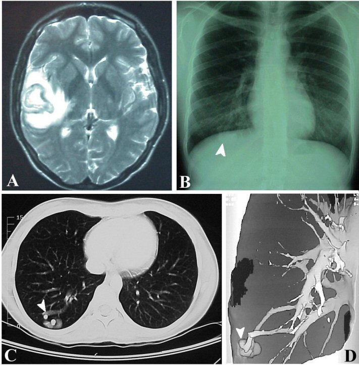
A 23-year-old woman presented with 3 days of headache followed by complex partial seizures.
(A) Axial T2-weighted MRI—right temporal mass with surrounding edema; (B) chest x-ray shows mass at right base; (C) axial chest CT—enhancing vascular mass at right base; (D) three-dimensional chest CT reconstruction demonstrates a right pulmonary arteriovenous fistula. Stereotactic aspiration of a large right temporal mass revealed purulent material with Gram-positive cocci (Streptococcus millieri). Chest radiograph and CT scan demonstrated a pulmonary arteriovenous fistula which resulted in the infection. (PAVF). PAVF are low resistance, abnormal connections between a pulmonary artery and distended vein. They may occur as an isolated entity or associated with hereditary hemorrhagic telangiectasia (Osler Weber Rendu syndrome). PAVF often present with neurologic symptoms, usually stroke, TIA, or brain abscess from the right to left shunt that bypasses the normal filtering action of the lungs. From David C. Preston and Barbara E. Shapiro, Pulmonary arteriovenous fistula and brain abscess. Neurology, Feb 2001; 56: 418.
Revised
06/10/04.
The Electronic Curriculum is copyrighted 1998, Case Western Reserve University
School of Medicine.