A 48 year-old woman developed headaches and difficulty with her vision. On examination, she had a bi-temporal field deficit.
![]()
![]()
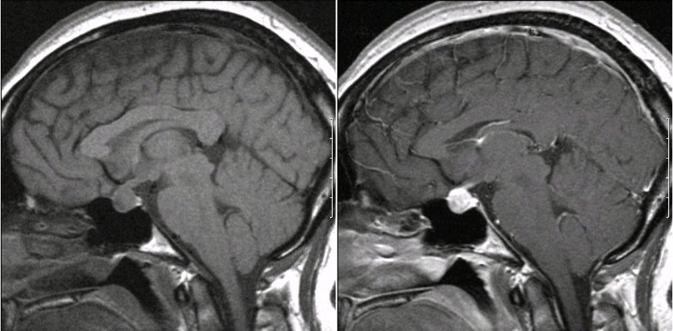
Sagittal MRIs. (Left) T1-weighed image; (Right) T1-weighted with gadolinium. Note the large enhancing mass which fills the sella. This is the typical appearance of a small pituitary macroadenoma that is growing beyond the sella and starting to compress the optic chiasm above. Macroadenoma was typically designated as tumors larger than 10 mm.
Revised
05/17/06.
The Electronic Curriculum is copyrighted 1998, Case Western Reserve University
School of Medicine.