An 82 year old woman was admitted for elective cardioversion for atrial fibrillation. After the procedure, she went to rehabilitation. She had a fall after slipping in the bathroom. After the fall, she was unable to walk as well, and had pain and bruising in her buttocks and down the back of her left leg. She was on coumadin. Upon re-admission, her INR was 6. Examination was normal except for weakness of left knee flexion and hip flexion, possibly limited by pain. Her patellar reflex on the right was 2+ and absent on the left. Ankle jerks were normal bilaterally. The medial part of her leg (as opposed to thigh) had decreased sensation.
![]()
![]()
![]()
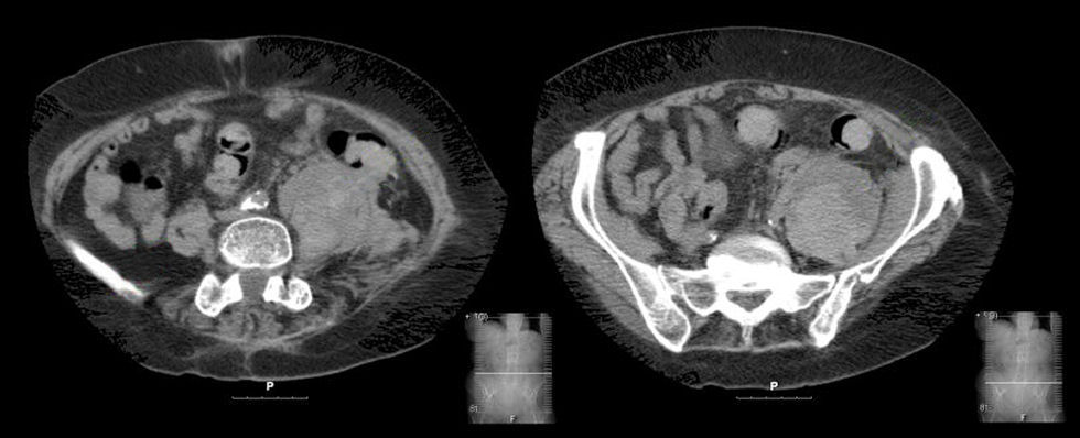
Axial CT Scan of the Abdomen and Pelvis: Note the large mass on the left. This is a retroperitoneal hematoma within the substance of the psoas muscle. Note the normal psoas muscle on the right. Spontaneous retroperitoneal hemorrhage into the psoas muscle is often associated with coagulopathies (e.g., excessive anticoagulation, hemophilia, etc.). The hematoma places pressure on the underlying lumbar plexus, resulting in weakness of hip flexion (iliopsoas), knee extension (quadriceps) and hip adduction (thigh adductors). The knee reflex is typically depressed. Sensory loss may be present over the anterior thigh, medial thigh, and/or medial calf. Pain often interferes with assessment of muscle strength.
Revised
04/26/06.
The Electronic Curriculum is copyrighted 1998, Case Western Reserve University
School of Medicine.