A 32 year old man presented with progressive numbness and pain radiating from the back to the anterior and medical thigh. of the face.
![]()
![]()
![]()
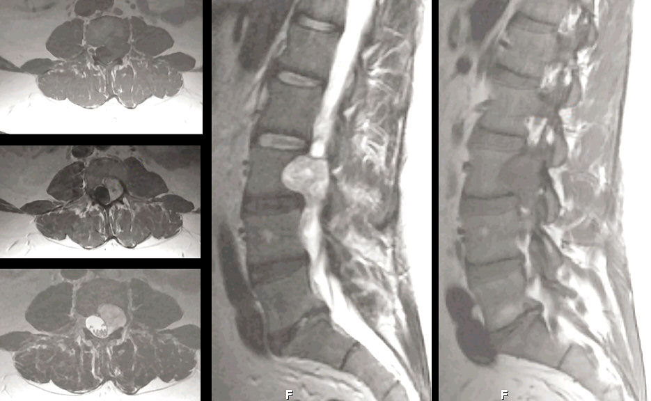
MRI Scans of the Lumbar Spine: (Left Top) Axial T1; (Left Middle) Axial T1 with Gadolinium; (Left Lower) Axial T2; (Middle) Mid-sagittal T2; (Right) Parasagital T1. Note the large nodular mass at the L3 level. On the axial scans, the lesion ehances with gadolinium. On the Left Lower image, one can clearly see that this lesion is extradural. On the parasagittal scan on the Right, note the normal nerve root with surrounding epidural fat. Contrast this to the lesion that is growing through and completely obliterating the foramen. At surgery, pathological examination revealed a schwannoma. Schwannomas are histologically benign tumors, seen along the course of peripheral nerves, nerve roots, and cranial nerves (especially CN V and VIII).
Revised
05/04/06.
The Electronic Curriculum is copyrighted 1998, Case Western Reserve University
School of Medicine.