A four-year old girl presented with a delay in motor milestones and seizures.
![]()
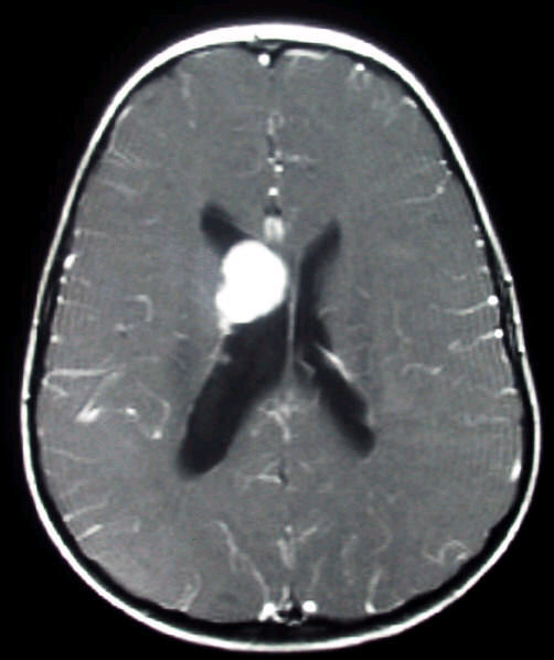
Axial MRI T1-weighted scan with Gadolinium. Note the large subependymal nodule on the right. This is a giant tuber in a patient with tuberous sclerosis (TS). In TS, several abnormalities may be present on brain imaging including, cortical tubers (hamartomas) subependymal nodules that usually calcify and are multiple and bilateral; and subependymal giant cell astrocytomas that are most frequently found at the foramen of Monro.
Revised
04/23/06.
The Electronic Curriculum is copyrighted 1998, Case Western Reserve University
School of Medicine.