A 52
year-old hypertensive man developed headache, nausea and vomiting, associated
with right sided numbness which slowly worsened over thirty minutes.
![]()
![]()
![]()
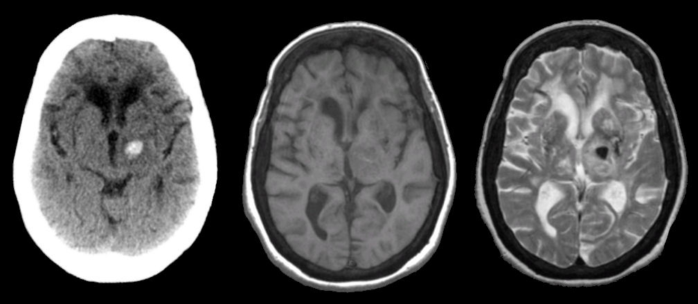
(Left) Axial CT scan:;
(Middle) T1-weighted image; (Right) T2-weighted image. Note the high density in
the left thalamus, indicating acute hemorrhage. On the T1-weighted image, the
abnormality is not well seen. However, note the decreased signal on the
T2-weighted image (representing deoxyhemoglobin. This is the MRI picture of an
acute/subacute stroke.
Revised
05/04/06.
The Electronic Curriculum is copyrighted 1998, Case Western Reserve University
School of Medicine.