A 5 year-old boy presented with epilepsy and mild developmental delay.
![]()
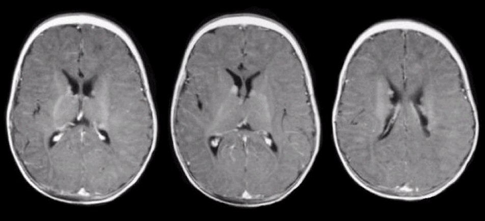
Axial MRI T1 weighted scan with Gadolinium. Note the numerous small enhancing lesions, most of which are subependymal near the ventricles. This is the typical appearance of tuberous sclerosis (TS). In TS, several abnormalities may be present on brain imaging including, cortical tubers (hamartomas) subependymal nodules that usually calcify and are multiple and bilateral; and subependymal giant cell astrocytomas that are most frequently found at the foramen of Monro.
Revised
05/19/06.
The Electronic Curriculum is copyrighted 1998, Case Western Reserve University
School of Medicine.