A 34year old woman presented with headaches that were suggestive of migraines.
![]()
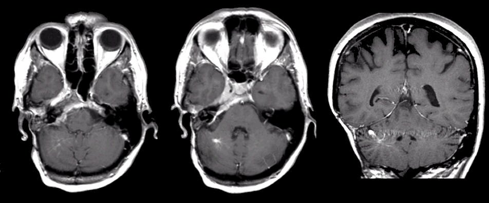
T1-weighted with gadolinium MRI scans. (Left and Middle) axial; (Middle) sagittal; (Right) coronal. Note the enhancing vascular structure adjacent in the right cerebellum. This is a venous angioma. Most angiomas are asymptomatic, as was the case in this patient.
Last Update:
3/14/06
The Electronic Curriculum is copyrighted 1998, Case Western
Reserve University
School of Medicine.