A 33 year old woman presented with a blunted affect and depression.
![]()
![]()
![]()
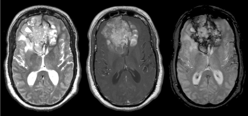
(Left) T2-weighted axial MRI; (Middle) T1-weighted with gadolinium axial image; (Right) Gradient echo axial image. Note the large enhancing mass in the frontal lobes. This pattern of a frontal tumor crossing over the midline via the corpus callosum to the contralateral frontal lobe is known as the butterfly pattern. On the gradient echo image, the dark signal identifies old blood. Biopsy showed glioblastoma multiforme.
Revised
05/07/06.
The Electronic Curriculum is copyrighted 1998, Case Western Reserve University
School of Medicine.