![]()
![]()
![]()
![]()
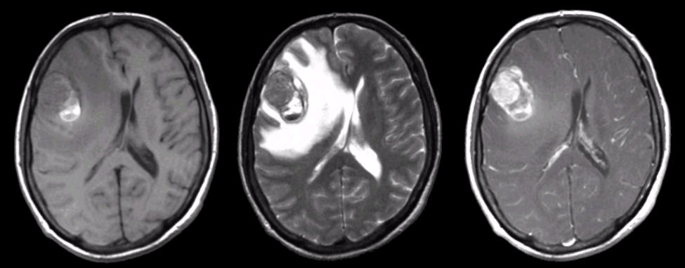
Axial MRI images: (Right) T1-weighted; (Middle) T2-weighted; (Right) T1-weighted with gadolinium. Note the large mass in the right frontal lobe that enhances with gadolinium (right image). Melanoma is one of the one common tumors to bleed. On the T1-weighted image, note the high signal which is a dark rim with a high signal on T2- weighted image. This is the MRI picture of subacute blood. The signal characteristics are those of intracellular methemoglobin.
Revised
05/16/06.
The Electronic Curriculum is copyrighted 1998, Case Western Reserve University
School of Medicine.