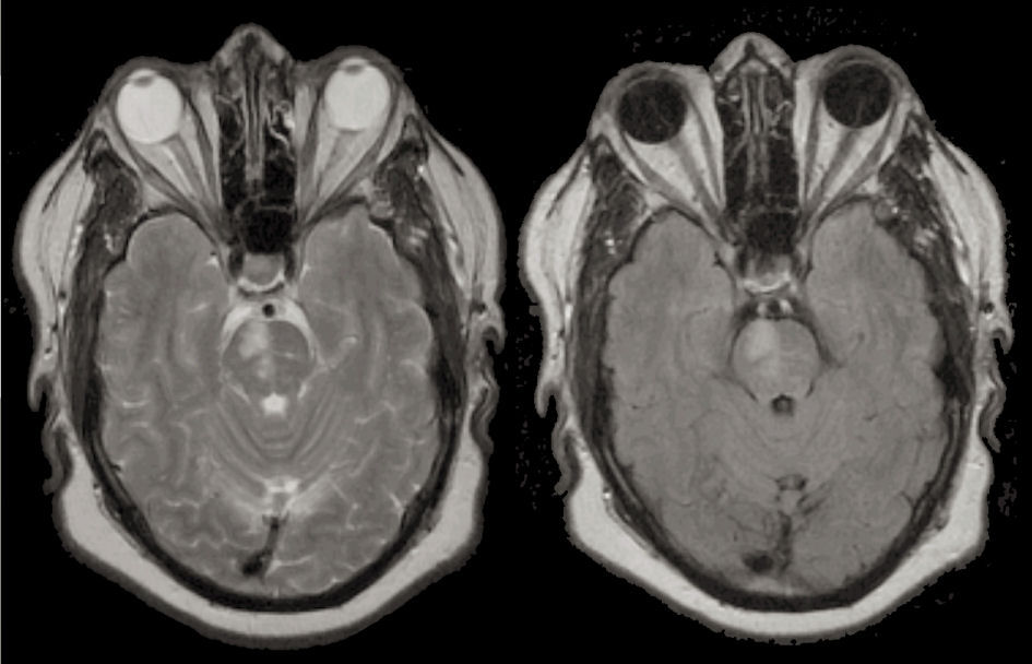
A 74 year old woman with hypertension and diabetes developed a left hemiparesis and left gaze preference.
![]()
![]()
(Left) T2 weighted axial MRI; (Right) Flair axial MRI. Note the high signal in the right pons. This lesion is in the distribution of one perforating branch off the basilar artery. Although occasionally associated with intrinsic basilar disease or an embolus to the basilar artery, this lesion usually represent the occlusion of one perforating basilar branch from the process of lipohyalinosis, which occurs as a result of aging, diabetes and hypertension. Also note the wide open basilar artery (flow void on the ventral pons).
Revised
05/10/06.
The Electronic Curriculum is copyrighted 1998, Case Western Reserve University
School of Medicine.