A 47 year-old woman presented with headaches.
![]()
![]()
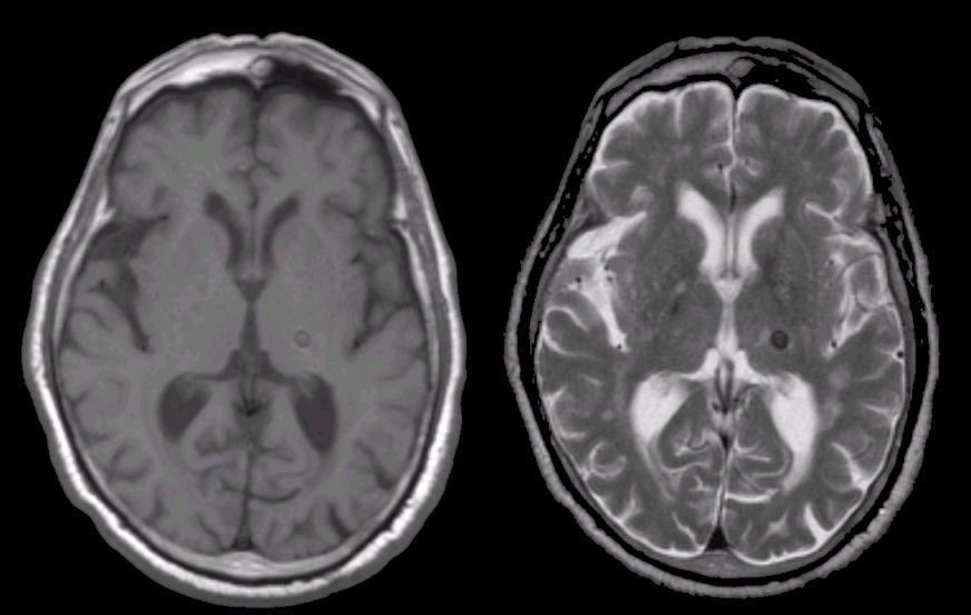
(Left) Axial T1 weighted MRI; (Right) T2 axial weighted MRI. Note the lesion which is dark on both T1 and T2 weighted images. This is consistent with hemosiderin, the residual from old blood. In this case, the old hemorrhage was caused by a cavernous angioma. To learn more, review the powerpoint slide show, Blood on MRI: Time-dependent Changes. In this case, the hemorrhage was due to hypertension.
Revised
05/02/06.
The Electronic Curriculum is copyrighted 1998, Case Western Reserve University
School of Medicine.