A 59 year-old man was evaluated for headaches. There was a history of an automobile accident ten years earlier, where he was in coma for several days after.
![]()
![]()
![]()
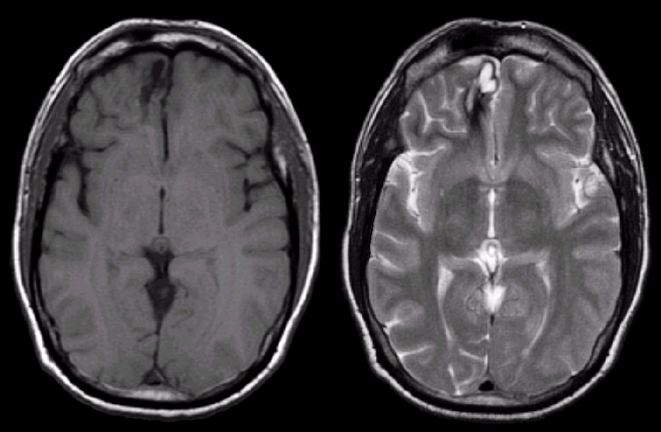
(Left) Axial T1 weighted MRI; (Right) T2 axial weighted MRI. Note the lesion in the right anterior frontal area which is dark on both T1 and T2 weighted images. This is consistent with hemosiderin, the residual from old blood. On the T2-weighted images, there is a small cystic area which is bright. To learn more, review the powerpoint slide show, Blood on MRI: Time-dependent Changes. In this case, the hemorrhage was due to hypertension.
Revised
05/02/06.
The Electronic Curriculum is copyrighted 1998, Case Western Reserve University
School of Medicine.