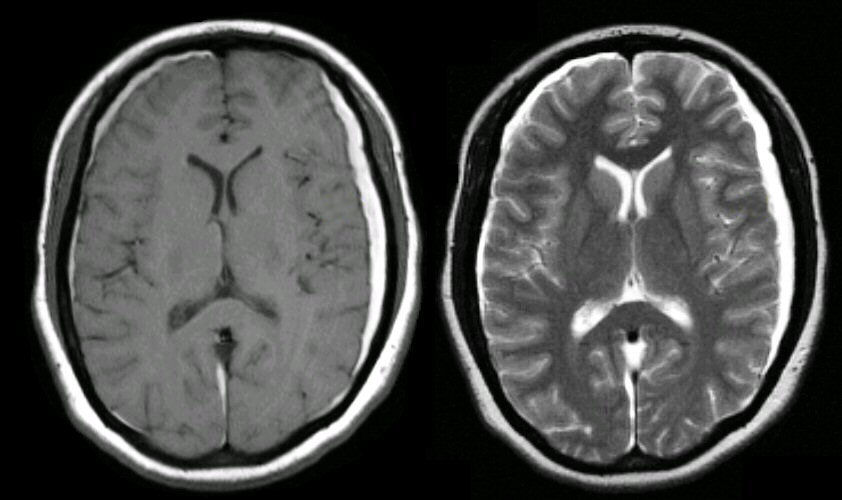
A 83 year old man developed confusion and difficulty walking after minor head trauma.
![]()
(Left) MRI axial T1 weighted image; (Right) MRI coronal T2 weighted image. Subdurals are recognized by their crescent shape overlying and compressing the brain. In the elderly, they can occur following minor trauma, affecting the so-called bridging veins between the dura and brain. Note the bilateral subdurals, more prominent on the left. On both T1 and T2 scans, the subdurals are bright, denoting extracelluar methemoglobuin.
Revised
05/06/06.
The Electronic Curriculum is copyrighted 1998, Case Western Reserve University
School of Medicine.