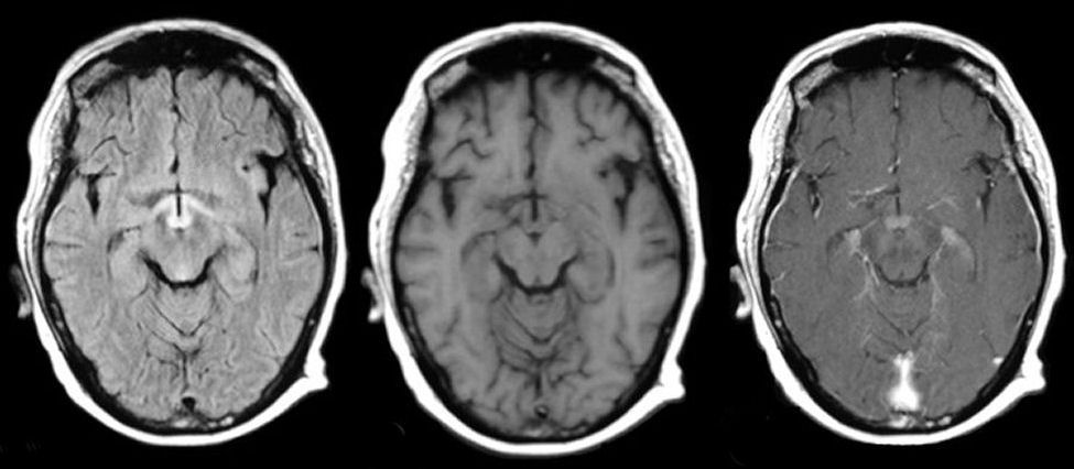
A 50 year old woman with intractable vomiting developed confusion, memory loss and loss of vision.
![]()
![]()
![]()
MRI scans: (Left) Axial Flair, (Middle) Axial T1-weighted, (Right) Axial T1-weighted with gadolinium. Note the enhancement of the mamillary bodies on the post-gadolinium scan and the prominent signal changes in the hypothalamus and mamillary bodies on the flair image. On the T2-weighted scan, note not only the involvement of the mamillary bodies, but alos the medial hypothalamus and optic tracts. Wernicke's encephalopathy occurs from thiamine deficiency, most classically seen with alcoholics, but also seen in poor nutrition of other causes. Note the prominent diffusion abnormalities and enhancement in the mamillary bodies, hypothalamus and optic tracts. This patient recovered completely several days after thiamine supplementation.
Revised
05/06/06.
The Electronic Curriculum is copyrighted 1998, Case Western Reserve University
School of Medicine.