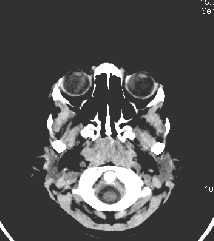
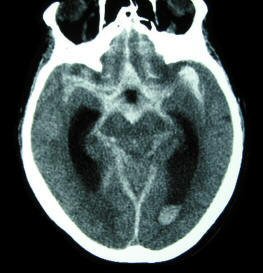
NEUROLOGIC INDICATIONS FOR CRANIAL CT
CT is most commonly indicated in the emergency room evaluation of acute head
trauma and acute neurologic dysfunction, primarily to look for suspected
intracranial or subarachnoid hemorrhage. Its extremely rapid acquisition time,
and sensitivity to detect hemorrhage makes it the modality of choice in the
acute setting time.
CT is easily available at nearly all institutions. Claustrophobia is not a
major issue, as it is in MRI.
In general, CT is useful in the
following conditions:
Vascular
Ischemic stroke (> 2 days old)
Hemorrhagic
stroke (acutely)
Tumor
Primary CNS and metastatic
Infection
Abscess
Hydrocephalus
Trauma
Epidural hematoma, subdural
hematoma, contusion, and skull fractures
|

 X-rays are applied in a circular motion with detectors on the opposite side of
the body. Body tissue slices (typically 1 cm) are mathematically reconstructed and
displayed on a gray scale matrix. The density of the tissue is in proportion to
the attenuation of the x-ray which pass through. Tissues like air and water have
little attenuation and are displayed as low densities (dark); whereas bone has
high attenuation and is displayed as a high density (bright) on CT. Among
pathologic conditions, high intensity lesions are often seen with freshly
clotted blood, hyperemia and with the use of contrast. Low intensity lesions include edema and necrosis.
X-rays are applied in a circular motion with detectors on the opposite side of
the body. Body tissue slices (typically 1 cm) are mathematically reconstructed and
displayed on a gray scale matrix. The density of the tissue is in proportion to
the attenuation of the x-ray which pass through. Tissues like air and water have
little attenuation and are displayed as low densities (dark); whereas bone has
high attenuation and is displayed as a high density (bright) on CT. Among
pathologic conditions, high intensity lesions are often seen with freshly
clotted blood, hyperemia and with the use of contrast. Low intensity lesions include edema and necrosis. 
